Costoclavicular Brachial Plexus Block

Indications: Hand, forearm, and elbow surgery, as well as upper arm surgery below the deltoid/shoulder if via a lateral skin incision.
Considerations: This is a variation on the more common, lateral-sagittal approach to the infraclavicular brachial plexus block. Instead of depositing local anesthetic into the lateral infraclavicular fossa with the ultrasound probe held perpendicular to the clavicle, the costoclavicular approach deposits local anesthetic slightly more medially, into the costoclavicular space, with the probe held parallel to the clavicle. In this location, the three cords of the plexus are consistently positioned clustered together just lateral to the artery – a nerve-artery visual relationship somewhat akin to the supraclavicular view, but performed inferior to the clavicle. In some pediatric patients, this approach may also offer more room to manipulate the needle.
Patient Position: Supine, with the neck tuned slightly to the contralateral side. Arm abducted to 90 degrees. A folded towel or other padding placed beneath the shoulder can aid in bringing the deep artery to a more superficial position.
Dose: 0.5 - 1.5 mg/kg of Bupivacaine or Ropivacaine (roughly 0.2 - 0.5 mL/kg). Larger doses can be used for bilateral blocks, as long as no more than 2.5 mg/kg is used in total. Rarely is more than a maximum volume of 20mL per block necessary.
Technique: Probe – Linear; Needle – In-plane
Begin with the probe directly over the mid-point of the clavicle and slide laterally and caudally to slip just off of its bony surface. Tilt the probe slightly so that the ultrasound beam points cephalad and allows the axillary artery, vein, and lateral cluster of cords to come into view. Optimize the view with very small clockwise and counterclockwise rotational probe adjustments until a cross-sectional view of the artery is achieved.
Needle entry is in-plane with a lateral to medial approach. Ideal needle tip positioning should be at the center of the 3 cords, in the gap between the lateral and posterior cords, but with care not to advance into the medical cord.
Occasionally the cephalic vein or thoracoacromial artery is encountered traversing your desired needle path. If this is the case, moving the probe medially and/or tilting more cephalad is often the key.
Coverage: Brachial Plexus – Roots level
Potential Complications:
Intravascular injection or injury
Pneumothorax / hemothorax (increased theoretical risk as compared to other brachial plexus blocks)
Hemidiaphragmatic weakness (lesser incidence than supraclavicular block, greater incidence than lateral-sagittal infraclavicular approach)
Bleeding / infection at needle insertion site
Patient Positioning and Probe Orientation


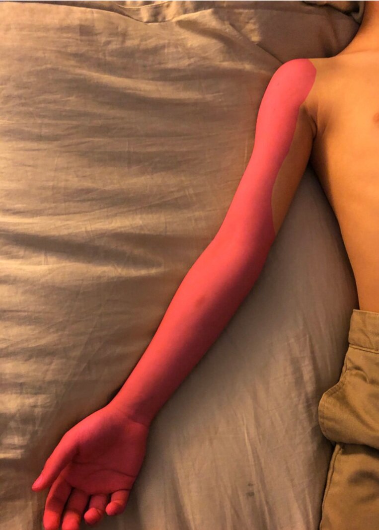
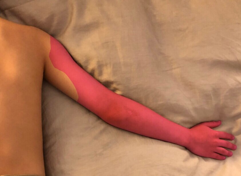
Expected Dermatomal Coverage
Ultrasound Images


The costoclavicular block was first described by in 2015 by Karmakar et al. As compared to the more traditional lateral-sagittal infraclavicular block technique, its benefits include: more superficial neurovascular landmarks, consistent cord location lateral to the artery, and cords approximated together in one cluster (Li et al. and Tinoco et al.). One drawback as compared to the lateral-sagittal approach is the increased potential for hemidiaphragmatic paralysis. Sivashanmugam et al found a lower incidence of this as compared to the supraclavicular block, but its incidence as compared to the lateral-sagittal infraclavicular block in pediatrics is not known. This may have important implications in patients with preexisting pulmonary disease or in those needing bilateral upper extremity surgery, such as for symbrachydactyly reconstruction.
As compared to the traditional infraclavicular approach in pediatric patients, Yayik et al describes a faster block performance time for the costoclavicular block. However, there was no difference in number of needle passes, needle visibility, block success, or block duration. Adult studies are conflicting on any benefit in onset time that the costoclavicular block has over the traditional approach. The difference may simply lie in the volume chosen in each of the studies.
Regarding technique, single, double, and even triple injection locations at the cords have been described. In awake adults, needle redirection to multiple locations has shown modest improvement in time to onset, though all approaches are effective. For our smallest of patients, most often under general anesthesia when the block is applied, the need for extremely rapid onset, is obviated. The author prefers the single injection technique for pediatric cases. This allows for as little needle manipulation as possible, in a block where the lung fields are more closely approximated to the needle than with any other brachial plexus block. This also takes into consideration the aberrant vascular anatomy Sivakumar et al. published caution on. If inadequate local anesthetic spread to all three cords via the single injection technique is noted on ultrasound visualization, only then should needle re-direction occur.
In the pediatric population, Carioca et al. note their groups’ preference for this block due to the superficial and compact brachial plexus positioning at this location. They showed a very low incidence of complications (a single arterial puncture with no long-term sequelae in 198 patients). Interestingly, their group noted a shift away from supraclavicular blocks and the traditional lateral-sagittal infraclavicular approach towards the costoclavicular block during the course of their 2-year study period.
Karmakar MK, Sala-Blanch X, Songthamwat B, Tsui BC. Benefits of the costoclavicular space for ultrasound-guided infraclavicular brachial plexus block: description of a costoclavicular approach. Reg Anesth Pain Med. 2015 May-Jun;40(3):287-8. doi: 10.1097/AAP.0000000000000232. PMID: 25899958.
Li JW, Songthamwat B, Samy W, Sala-Blanch X, Karmakar MK. Ultrasound-Guided Costoclavicular Brachial Plexus Block: Sonoanatomy, Technique, and Block Dynamics. Reg Anesth Pain Med. 2017 Mar/Apr;42(2):233-240. doi: 10.1097/AAP.0000000000000566. PMID: 28157792.
Tinoco J, Eloy A, Regufe R. Costoclavicular brachial plexus block: A review of current evidence. Rev Esp Anestesiol Reanim (Engl Ed). 2022 Dec;69(10):649-653. doi: 10.1016/j.redare.2022.10.004. Epub 2022 Nov 4. PMID: 36344407.
Sivashanmugam T, Maurya I, Kumar N, Karmakar MK. Ipsilateral hemidiaphragmatic paresis after a supraclavicular and costoclavicular brachial plexus block: A randomised observer blinded study. Eur J Anaesthesiol. 2019 Oct;36(10):787-795. doi: 10.1097/EJA.0000000000001069. PMID: 31397702.
Yayik AM, Cesur S, Ozturk F, Celik EC, Naldan ME, Ahiskalioglu A. Comparison of the lateral sagittal and costoclavicular approaches for ultrasound-guided infraclavicular block in pediatric patients: a prospective randomized study. Braz J Anesthesiol. 2021 Jun 6:S0104-0014(21)00224-4. doi: 10.1016/j.bjane.2021.05.005. Epub ahead of print. PMID: 34090921.
Sivakumar RK, Areeruk P, Karmakar MK. Aberrant vascular anatomy at the costoclavicular space: a word of caution for costoclavicular brachial plexus block. Reg Anesth Pain Med. 2021 Jan;46(1):95-96. doi: 10.1136/rapm-2020-101498. Epub 2020 Apr 20. PMID: 32317294.
Carioca F, Silva M, Bispo C, Mafra J, Cenicante T. Costoclavicular brachial plexus block in paediatric anaesthesia: A retrospective pilot study. J Clin Anesth. 2021 May;69:110113. doi: 10.1016/j.jclinane.2020.110113. Epub 2020 Nov 13. PMID: 33190038.