Allen and Barbeau Tests

Things that we are supposed to do every day in the course of our adult lives: wake up early, wash behind your ears, have a balanced breakfast, floss, brew a cup of organically grown, fair-trade, locally roasted coffee!
I’m sure many of us can still hear our parents voices reminding us to follow this internal daily checklist to a T.
Lest you think these daily tasks are only reserved for life outside the OR, think again! As has already been discussed elsewhere, safety is a top priority of our profession. Machine functionality, check. Pilot balloon functioning, check. Laryngoscope battery, check. Name band, check. Correct side site marked, check.
Allen test, check?
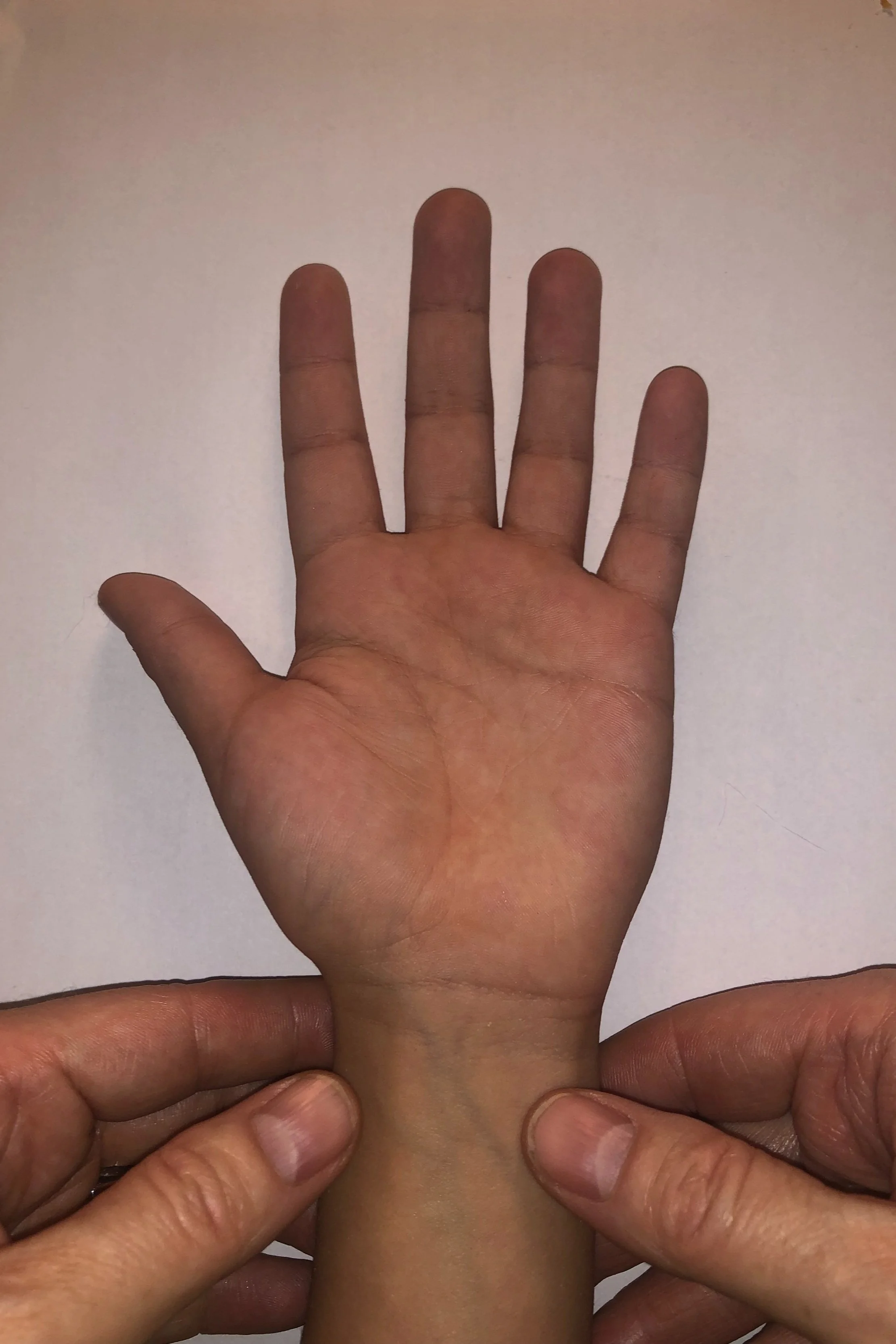
Allen Test
The Allen Test was first described in 1929 by Edgar V. Allen, at the time an Internal Medicine Fellow at the Mayo Clinic. It was designed to elicit otherwise undiagnosed occlusive lesions in the arteries distal to the wrist, namely Thromboangiitis Obliterans (aka Buerger's Disease), a smoking-related condition that results in blood clot formation in small and medium-sized arteries. In the originally described technique, one occluded the non-suspect artery at the wrist, along with the patient making a fist for one minute. This would effectively drain the blood from the hand. After relaxation of the hand (but maintaining pressure on the compressed artery), “in individuals with an intact arterial tree the pallow is quickly replaced by rubor of a higher degree than normal, which gradually fades to the normal color.” If the uncompressed artery is diseased, “pallor is maintained for a variable period, due to the obstruction to arterial inflow in the two main channels.”
In 1952, Irving Wright described a modified version of the Allen test (MAT) that has since largely supplanted the original method. In the modified version, one is explicitly looking for ulnar pathology (or patent collateral circulation) prior to radial artery cannulation. As such, both the radial and ulnar arteries are compressed while the patient makes a fist. After blanching of the hand, ulnar artery compression is released. In the absence of ulnar pathology, normal color should return within 2-5s, though the definition of an “abnormal” Allen Test is somewhat ill-defined, as is it’s reliability. To date, no study has demonstrated a relationship between the time to maximal blushing using the MAT and ischemic symptoms or complications to the hand. Nevertheless, the MAT is still routinely advocated to avoid acute and long-term ischemia of the hand in cases of radial artery occlusion.
Barone et al published a review article in 2006 looking at all the available literature at the time. Heterogeneous as the studies were, the researchers found some startling things. First, the definition of “abnormal” is quite variable. Some sources say a refill time > 5s, while others say ≥ 15s should be considered abnormal. In one study looking at inter-observer reliability, four physicians examined 200 hands of healthy subjects. The four physicians never all agreed that any one test was abnormal. Not only is the definition of abnormal variable, but what constitutes abnormal can’t even be agreed upon during physical examination! In a paper comparing the MAT to Doppler, the MAT was accurate only 80% of the time when the abnormal limit was >5s. The authors concluded that the Allen test is “unreliable for deciding which patients could safely undergo radial artery grafting for coronary artery bypass surgery.” In fact, even patients with a normal Allen Test are still at risk for ischemia.
Modified Allen Test


Transradial Access (TRA) has also become more popular in the adult interventional cardiology realm.I can still remember being an intern almost 15 years ago and listening to the cardiology fellow speak in hushed tones that Dr. So-and-So did their caths through the radial artery. Although radial artery occlusion (RAO) has been documented in up to 30% of patients after transradial catheterization, it is a common clinical observation that RAO remains asymptomatic in the large majority of patients. To date, no hand or finger amputation has been reported after TRA for diagnostic angiography or intervention, even though TRA has been used worldwide for more than 20 years.
One limitation that also bears mentioning, to properly perform the MAT one typically requires a cooperative patient. However, one can perform the MAT in uncooperative patients (read infants and anesthetized patients) by elevating the hand and compressing the radial and ulnar arteries until the hand blanches.
Barbeau Test
An alternative screening modality is the Barbeau Test, one that is potentially both more sensitive, as well as less subjective. It can also be done in uncooperative and sedated patients, i.e. most of our pediatric patients.
To perform the Barbeau Test, one first places a pulse oximeter ideally on the thumb. The ulnar artery is often dominant in the hand circulation, but the thenar eminence and the thumb are predominantly dependent on the radial artery, especially when the palmar arches are incomplete. If the thumb cannot accommodate the pulse oximeter, placing it on the 1st finger may suffice (as it is also largely supplied by the radial artery). Ensure an adequate pulse ox waveform. Compress both arteries until you see waveform flattening. Release the artery you don’t plan on cannulating to determine if adequate collateral circulation exists. If you plan on cannulating the radial, release the ulnar. If you plan on cannulating the ulnar, release the radial. The authors categorized their results into 4 types of ulnopalmar arch patency (A-D), with A showing no interruption in perfusion and D showing absent plethysmography even after 2 minutes. Similar to the MAT, no relationship to plethysmography/oximetry and ischemic symptoms or complications after radial artery cannulation has been found. Due to the increased sensitivity of the Barbeau Test, it may be that those with a concerning result are weeded out. However, an abstract presented by the same group in over 7,000 patients, regardless of the Barbeau Test result, had zero cases of hand ischemia.
In another application of the Barbeau Test, the PROPHET Study evaluated radial artery patency after catheterization (co-authored by Dr So-and-So no less!). Patients were randomized to either conventional pressure after catheter removal vs applying pressure in conjunction with the Barbeau test (maintaining radial artery patency while holding pressure). They found that maintaining radial artery patency while achieving hemostasis is highly effective in reducing radial artery occlusion after radial access. The authors recommended that guided compression (with the Barbeau Test) after catheter removal should be performed to prevent radial artery occlusion.

Neither the MAT or Barbeau Test has been studied to any great extent in the pediatric population, so one cannot draw any definitive conclusions on the predictive ability of either modality (much the same as in the adult population!). Thankfully, while undeniably devastating, the incidence of ischemia following arterial catheterization seems to be quite low. In adults, the incidence of severe or permanent ischaemia has been observed in 0.2–0.75% of adults. In a paper hot off the presses, Gleich et al describe their single institution pediatric experience. In their evaluation of over 5,000 arterial lines (66% radial, 29.7% femoral), they had 8 vascular complications (1 for bleeding, 7 for ischemia), all of which occurred in the femoral artery. Vascular complications occurred earlier in a patient’s course at median 1.5 days, with neonates and infants having the highest risk. While the number of attempts are not listed, ultrasound was used in 86% of the ischemic femoral lines. Based on their data, the incidence of severe ischemia in kids is roughly around 0.14%. The authors noted that there were no complications in distal arterial cannulation sites, including more than 3,395 radial cannulations! Secondary to these results, the authors have since changed their practice and preferentially place radial or ulnar arterial lines in all age groups if possible. Their results are very similar to Kayssi et al who evaluated pediatric patients for acute limb ischemia over a 13 year period. They found that 91% of acute limb ischemia cases were secondary to catheterization, of which 94% occured in the lower extremity. Thankfully, 94% of those patients were treated with anticoagulation alone, and only 2% ultimately required surgical intervention.
Since we may not be able to selectively screen for high risk patients, we might instead consider focusing on things we have some control over, notably early recognition of ischemia and modifying risk factors. Some clinical signs of subclinical ischemia are reduced temperature and weak peripheral ipsilateral pulses. The features of limb-threatening ischemia include the presence of skin mottling, paralysis, and absent limb pulses with no capillary refill. Risk factors for ischemia following catheterization include prolonged catheterization, distal vs proximal location, catheter size and composition, age < 3 years, hypotension, spasm secondary to repeated arterial puncture, ≥ 3 catheterization attempts, pre-existing arterial disease and sustained external pressure to achieve hemostasis after catheter removal. Though rarely due to the catheter by itself, the presence of shock compounds the rate of complications and ischemia.
With these things in mind, one should attempt to use the smallest viable catheter, preferentially placed in a distal artery. It is also recommended that one should advance the needle at less of an acute angle during placement (presumably to increase first attempt success). Ultrasound-guidance should also be heavily encouraged. Monitor extremities for signs of vascular compromise frequently: examine tips of fingers or toes for peripheral catheters, the lower extremities including buttocks for UACs. Remove catheters as soon as medically feasible, or if there are signs of vascular compromise or suspicion of thrombosis. Perhaps also consider the Barbeau Test when you pull the catheter and are applying pressure for site hemostasis. Some would also suggest alternative arterial cannulation sites with more robust collaterals, e.g. posterior tibial or dorsalis pedis. At least one source even advocates for the temporal artery!
Not everyone agrees.
Honestly, I still shudder a bit with scalp IV’s. I might just consider going without arterial access if I found myself down to cannulating the temporal artery. But that may just be me.
Allen, E.V., 1929. Thromboangiitis obliterans: methods of diagnosis of chronic occlusive arterial lesions distal to the wrist with illustrative cases. Am J. Med. Sci, 17, pp.237-244.
Wright IS (1952) Vascular diseases in clinical practice. 2nd ed. Chicago: The Year Book Publishers Inc
Ejrup, B., Fischer, B. and Wright, I.S., 1966. Clinical evaluation of blood flow to the hand: the false-positive Allen test. Circulation, 33(5), pp.778-780.
Fuhrman, T.M., Pippin, W.D., Talmage, L.A. and Reilley, T.E., 1992. Evaluation of collateral circulation of the hand. Journal of clinical monitoring, 8(1), pp.28-32.
Barone, J.E. and Madlinger, R.V., 2006. Should an Allen test be performed before radial artery cannulation?. Journal of Trauma and Acute Care Surgery, 61(2), pp.468-470.
Vu-Rose, T., Ebramzadeh, E., Lane, C.S. and Kuschner, S.H., 1997. The Allen test. A study of inter-observer reliability. Bulletin (Hospital for Joint Diseases (New York, NY)), 56(2), pp.99-101.
Jarvis, M.A., Jarvis, C.L., Jones, P.R. and Spyt, T.J., 2000. Reliability of Allen’s test in selection of patients for radial artery harvest. The Annals of thoracic surgery, 70(4), pp.1362-1365.
Toursarkissian, B., Mejia, A., Smilanich, R.P., Shireman, P.K. and Sykes, M.T., 2001. Changing pattern of access site complications with the use of percutaneous closure devices. Vascular surgery, 35(3), pp.203-206.
Nunoo-Mensah, J., 1998. An unexpected complication after harvesting of the radial artery for coronary artery bypass grafting. The Annals of thoracic surgery, 66(3), pp.929-931.
Barbeau, G.R., Arsenault, F., Dugas, L., Simard, S. and Larivière, M.M., 2004. Evaluation of the ulnopalmar arterial arches with pulse oximetry and plethysmography: comparison with the Allen's test in 1010 patients. American heart journal, 147(3), pp.489-493.
Barbeau, G.R., Gleeton, O. and Roy, L., 1999. Transradial approach for coronary interventions: Procedural results and vascular complications of a series of 7049 procedures. Circulation, 100, pp.1-306.
Green, J.A. and Tonkin, M.A., 1999. Ischaemia of the hand in infants following radial or ulnar artery catheterisation. Hand Surgery, 4(02), pp.151-157.
Gleich, S.J., Wong, A.V., Handlogten, K.S., Thum, D.E. and Nemergut, M.E., 2021. Major short-term complications of arterial cannulation for monitoring in children. Anesthesiology, 134(1), pp.26-34.
Kayssi, A., Shaikh, F., Roche-Nagle, G., Brandao, L.R., Williams, S.A. and Rubin, B.B., 2014. Management of acute limb ischemia in the pediatric population. Journal of vascular surgery, 60(1), pp.106-110.
Shiloh, A.L., Savel, R.H., Paulin, L.M. and Eisen, L.A., 2011. Ultrasound-guided catheterization of the radial artery: a systematic review and meta-analysis of randomized controlled trials. Chest, 139(3), pp.524-529.
Schwemmer, U., Arzet, H.A., Trautner, H., Rauch, S., Roewer, N. and Greim, C.A., 2006. Ultrasound-guided arterial cannulation in infants improves success rate. European journal of anaesthesiology, 23(6), pp.476-480.
Mansfield PB, Gazzaniga AB, Litwin SB. Management of arterial injuries related to cardiac catheterization in children and young adults. Circulation. 1970;42(3):501-507
Baker RJ, Chunprapaph B, Nyhus LM. Severe ischemia of the hand following radial artery catheterization. Surgery. 1976 Oct;80(4):449-57.
Ramasethu, J., 2008. Complications of vascular catheters in the neonatal intensive care unit. Clinics in perinatology, 35(1), pp.199-222.