Popliteal Sciatic Nerve Block

Indications: Foot/ankle surgery
Usually in combination with Saphenous/Adductor Canal Nerve Block
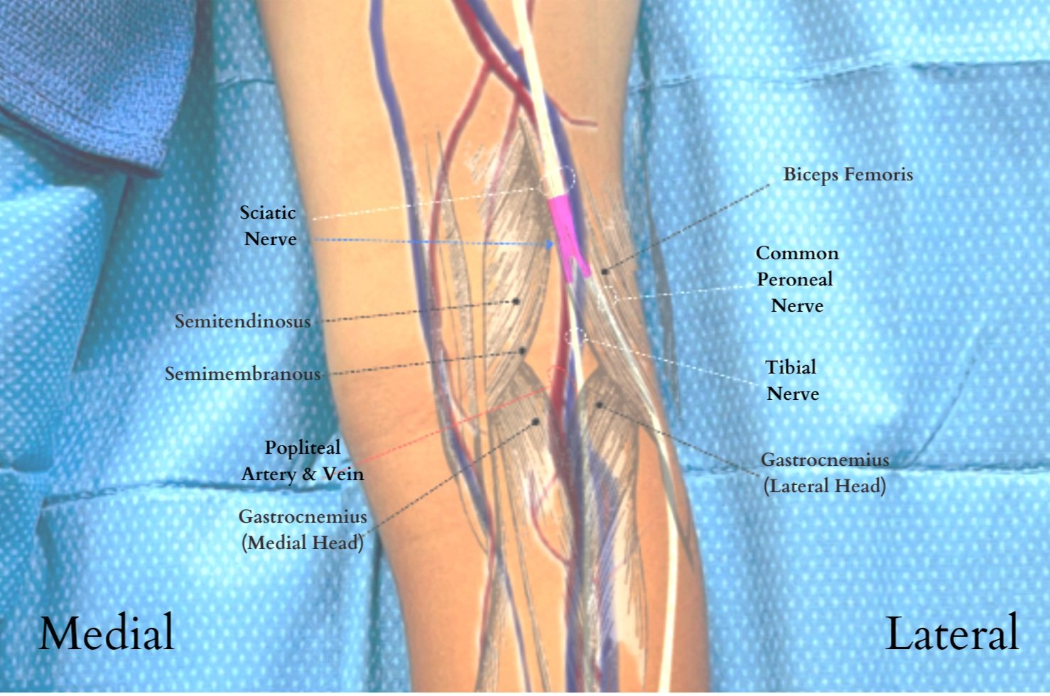
Anatomical Landmarks for Popliteal Block:
Lateral border: biceps femoris
Medial border: semimembranosus & semitendinosus muscles
Sciatic nerve lies deep to the muscles
Popliteal artery typically the deepest and most medial structure
Technique: Probe- Linear; Needle—in-plane
Can be done in either the supine, lateral, or prone position. Based on ease of access, many prefer having the patient lie prone. First, identify the popliteal artery/vein to locate the tibial nerve and scan cephalad to where the tibial nerve joins the common peroneal nerve to form the sciatic nerve (the tibial nerve will diverge from the popliteal vessels to join the common peroneal nerve)
Local anesthetic is injected at the level of the single sciatic nerve using an in-plane approach with the nerve viewed in cross section. If single approach does not result in circumferential spread, the first injection should be deep to the nerve with the second injection superficial. This way the first injection does not distort the image for the second injection.
Can also be done out-of-plane, though our preference is to perform in-plane to be able to identify the needle tip throughout the entirety of the procedure.
Dose: 0.5 - 1.5 mg/kg of Bupivacaine or Ropivacaine (roughly 0.15 - 0.3 mL/kg).
Potential Complications:
General Complications
Infection
Bleeding/hematoma
Damage to surrounding structures
Intravascular injection
Patient Positioning & Probe Orientation


Ultrasound Images & Block Video
Sc: Sciatic Nerve; CP: Common Peroneal Nerve; T: Tibial Nerve; BF: Biceps Femoris Muscle; SM: Semimembranosus Muscle
Wejjakul et al, 2021: randomized control trial looking at popliteal nerve block versus local surgical site infiltration for children ages 1-15 years old presenting for foot and ankle surgery. In younger age group (1-6 years old), no statistical significance in analgesia. However, in the older age group (7-15 years old), superior analgesia in the popliteal nerve block group .
Oberndorfer et al, 2007: prospective randomized trial comparing ultrasound guided versus nerve stimulator guided sciatic and femoral nerve blocks for children up to 8 years old for surgery of the lower extremity. All blocks done under general anesthesia. Sciatic (subgluteal or popliteal) +/- femoral nerve block as appropriate for surgical procedure, randomized to either ultrasound or nerve stimulator. Variable volume of local anesthetic (levobupivacaine 0.5%) for ultrasound guided blocks, local injected until seen to surround the nerve on ultrasound imaging. In contrast, set volume (0.3 ml/kg) of local anesthetic (levobupivacaine 0.5%) used for nerve stimulator guided blocks. Block failure defined by 15% heart rate increase from baseline. Duration of block defined by time from block to first analgesic administration. 2 block failures in nerve stimulator group, 0 in ultrasound group. Less local required for ultrasound group (p<0.001) for both sciatic nerve block (0.2 versus 0.3 ml/kg) and femoral nerve block (0.15 versus 0.3 ml/kg). Mean duration of block significantly longer (p<0.001) for ultrasound versus nerve stimulator group (mean 508 versus 335 min).
Roberts S. Ultrasonographic guidance in pediatric regional anesthesia. Part 2: techniques. Paediatr Anaesth. 2006 Nov;16(11):1112-24. doi: 10.1111/j.1460-9592.2006.02021.x. Erratum in: Paediatr Anaesth. 2006 Dec;16(12):1305-6. PMID: 17040299.
Tsui B, Suresh S. Ultrasound imaging for regional anesthesia in infants, children, and adolescents: a review of current literature and its application in the practice of extremity and trunk blocks. Anesthesiology. 2010 Feb;112(2):473-92. doi: 10.1097/ALN.0b013e3181c5dfd7. PMID: 20068455.
Wejjakul, W., Tangwiwat, S., Pangthipampai, P., Halilamien, P. and Eamsobhana, P., 2022. Does ultrasound-guided popliteal-sciatic nerve block have superior pain control in pediatric foot and ankle surgery? A randomized control trial. Journal of Orthopaedic Science, 27(4), pp.844-849.
Oberndorfer, U., Marhofer, P., Bösenberg, A., Willschke, H., Felfernig, M., Weintraud, M., Kapral, S. and Kettner, S.C., 2007. Ultrasonographic guidance for sciatic and femoral nerve blocks in children. British journal of anaesthesia, 98(6), pp.797-801.